
Pathology moved from glass slides under a microscope to whole-slide imaging on a screen. Artificial intelligence-driven tools now read those digital images, flag suspicious regions, and quantify features in seconds. This shift powers AI in pathology cancer detection across tumor types, sharpening both diagnosis and prognosis for patients.
Where AI Fits in the Digital Pathology Workflow
This article maps where AI enters the diagnostic pipeline, and where it does not. It covers foundation models, breast cancer histology grading, and biomarker prediction. It sets aside radiology, sequencing pipelines, and lab robotics.
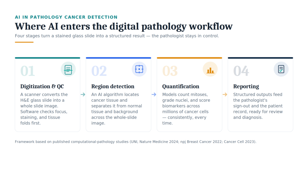
Digital pathology begins when a scanner turns a stained glass slide into a whole slide image. From there, AI in pathology cancer detection enters at four points:
- Digitization and quality control. Software checks focus, staining, and tissue folds before any image analysis starts.
- Region detection. An AI algorithm locates cancer tissue and separates it from normal tissue and background.
- Quantification. Models count mitoses, grade nuclei, and score biomarkers across millions of cancer cells.
- Reporting. Structured outputs feed the pathologist’s sign-out and the patient record.
Each stage produces measurable pathology data. Clinical directors can therefore target investment where returns are clearest. For most labs, region detection and quantification deliver the fastest payback. Founders, meanwhile, should note that these AI applications sit inside existing digital pathology platforms rather than replacing them. The pathologist stays in control. AI handles the repetitive measurement that slows a manual read. This division of labor frames the three examples below, each drawn from a peer-reviewed study.
Why AI Now Matters for Cancer Diagnostics
Three pressures push labs toward automation. Rising cancer cases, a shrinking pathologist workforce, and inter-observer variability all create delays in cancer diagnosis. AI in pathology cancer detection addresses these challenges with speed and consistency. These tools augment the expert; they do not replace one. The studies below show how AI applications revolutionize cancer detection and deliver more consistent cancer diagnoses.
Foundation Models Trained on Cancer Histology Slides
A pathology foundation model learns general tissue features from millions of images, then adapts to many tasks with little extra labeling. UNI, published in Nature Medicine [1], is a clear foundation model for computational pathology.
The team pretrained UNI on more than 100 million images drawn from over 100,000 diagnostic H&E-stained whole-slide images. That corpus spans 20 major tissue types and 77 terabytes of data. This stage required no labels. The model then faced 34 computational pathology tasks of varying difficulty.
Results across those tasks beat previous state-of-the-art encoders. UNI also classified up to 108 cancer subtypes in the OncoTree system, including breast, renal, and lung categories. One adaptable model handled tissue classification, slide classification, and cancer histology retrieval.
For a lab covering several cancer types, this matters. A single foundation model replaces a dozen narrow, single-purpose tools. Each new task needs fewer labeled slides, so cost falls and deployment speeds up. That efficiency explains why foundation models now anchor most new digital pathology research.
Deep Learning for Breast Cancer Histology Grading
Breast cancer grading rests on the Nottingham system: mitotic count, nuclear pleomorphism, and tubule formation. Each score is subjective, so two pathologists can disagree. A deep learning study in npj Breast Cancer [2] tackled mitosis detection in breast cancer alongside the other two components at once.
The researchers built a separate model per feature, then tested them on TCGA breast cancer tissue: 685 whole-slide images from 662 cases. Against a three-pathologist reference, slide-level agreement was strong:
| Grading feature | Model vs. pathologists (weighted kappa) |
|---|---|
| Mitotic count | 0.81 |
| Tubule formation | 0.75 |
| Nuclear pleomorphism | 0.48 |
More telling, model-to-pathologist agreement often beat agreement between the pathologists themselves. For mitotic count, the model scored 0.64 against a 0.56 human baseline; for tubule formation, 0.68 against 0.55. The mitosis model reached an F1 of 0.60 while counting individual tumor cells.
Consistent scores carry a prognostic payoff. The AI grade predicted outcomes on par with expert review, and its mitotic count tracked Ki-67 more tightly than the pathologists’ count did. For breast pathology, that reproducibility is where digital pathology and AI earn their place in diagnosis and prognosis, and improve cancer prognosis at scale.
AI-Predicted Biomarkers From Routine Tissue Slides
Some biomarkers show up directly in H&E pathology slides, with no separate molecular assay. Microsatellite instability (MSI) guides immunotherapy in colorectal cancer, yet lab testing adds days and cost. A transformer-based AI model can predict it from the slide alone.
Wagner and colleagues trained such a model on more than 13,000 patients across 16 cohorts. The system reads cancer biomarkers directly from cancer on H&E-stained whole-slide images.
Reported performance on held-out data:
- MSI: AUROC 0.97 on resections, 0.92 on biopsies, and 0.86 on an external cohort.
- Sensitivity: 0.99, with a negative predictive value above 0.99 on resections.
That sensitivity matters. A near-perfect negative predictive value lets a lab rule out cases up front, then send only flagged slides for confirmatory testing. This AI application reaches clinical-grade computational pathology on biopsy tissue, a long-standing weak point. The result is faster cancer diagnostics and lower molecular-testing spend, with no new equipment.
Future Directions for Digital Pathology and AI
AI-driven digital pathology keeps advancing. So where does AI in pathology cancer detection go next? Four directions stand out.
- Multimodal models. The strongest systems will fuse cancer histology with genomics, radiology, and clinical notes. This extends earlier work that predicted mutations from non-small cell lung cancer histopathology images using deep learning.
- Regulation. More tools will clear FDA and CE pathways. Gleason grading of prostate cancer on H&E-stained whole-slide images and MSI detection already show what approved products look like.
- Integration. Vendors are wiring models into digital pathology platforms and hospital systems, so results reach the pathologist without extra clicks.
- Validation. Honest gaps remain. Models must generalize across diverse populations, explain their predictions, and earn clinician trust.
These future directions in AI-driven cancer care depend on shared pathology data and prospective clinical trial evidence. Retrospective accuracy is not enough. Cancer research now needs trials that measure real effects on clinical decision-making and patient outcomes. Gastric, colorectal, breast, and prostate programs all sit on the same path. For founders and clinical leads, the roadmap is concrete, and the open problems look solvable rather than distant.
Partner With Us to Integrate AI Into Your Pathology Workflow
Ready to bring AI in pathology cancer detection into your lab? We help clinical directors and health-tech teams adopt AI across pathology workflows, from vendor selection to validation and rollout. Let’s turn these digital pathology solutions into results your pathology labs can trust. Contact us to scope your first project.
References
- Chen, Richard J., et al. “Towards a general-purpose foundation model for computational pathology.” Nature medicine 30.3 (2024): 850-862.
- Jaroensri, Ronnachai, et al. “Deep learning models for histologic grading of breast cancer and association with disease prognosis.” NPJ Breast cancer 8.1 (2022): 113.
- Wagner, Sophia J., et al. “Transformer-based biomarker prediction from colorectal cancer histology: A large-scale multicentric study.” Cancer cell 41.9 (2023): 1650-1661.