
A standard electrocardiogram takes five minutes and costs almost nothing. Now the same test can flag hidden heart problems early. This guide explains how AI detects heart disease from ECG, turning a routine strip into a screening tool. An AI model reads patterns that once needed an echo or MRI. Below, we cover four concrete uses.
What AI Adds to ECG Analysis
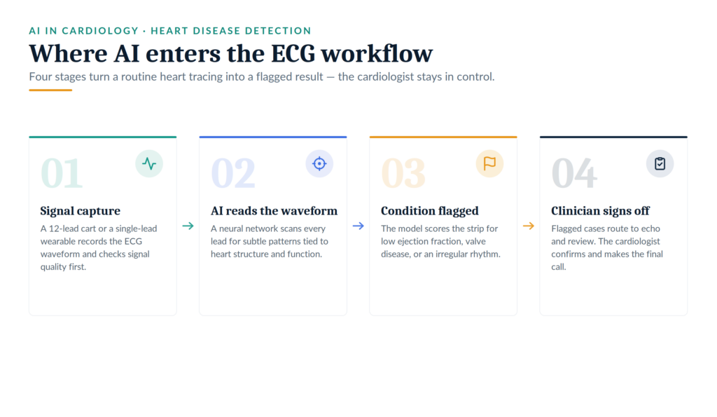
For a century, clinicians have read the electrocardiogram for rhythm and rate. Modern ECG analysis goes further. A neural network studies the full ECG waveform across all ECG leads, then links subtle ECG features to the heart’s structure and function.
Traditional ECG interpretation spots obvious abnormalities. Machine learning catches signals too faint for the human eye. The model trains on millions of paired ECG recordings and echocardiograms, so it maps ECG patterns to conditions such as weak pumping or restricted blood flow.
Two signal types feed these systems:
- 12-lead ECG from a clinic cart, rich with detail.
- Single-lead ECG from a watch or patch, simpler but portable.
An AI algorithm trained on this ECG data answers concrete clinical questions. Does this heart pump normally? Is heart function slipping? Should this patient get an echo? Those answers show how AI detects heart disease from ECG at scale.
One point matters for adoption. These tools support the clinician; they do not replace judgment. The cardiologist still signs off. Good AI explainability helps too, since a saliency map can highlight which part of the 12-lead ECG waveform drove the prediction. That transparency builds trust in clinical practice.
Why an AI Tool for the Electrocardiogram Matters Now
Heart disease often stays silent. Many patients have no symptoms until the disease is advanced. An AI tool turns a five-minute electrocardiogram into an early-warning system. The American Heart Association and regulators now back this shift. These three uses show how AI detects heart disease from ECG in real cardiovascular care.
Screening for Structural Heart Disease and Valve Disease in Primary Care
A family doctor records a routine ECG. The patient feels fine. Yet the model flags a high chance of aortic stenosis, which echocardiography later confirms. That workflow is now real.
Researchers built a deep-learning algorithm for detecting aortic stenosis using electrocardiography, published in J Am Heart Assoc [1]. The model learned from large sets of paired ECGs and echocardiograms. It then scored new strips for early aortic disease.
Performance held up on outside data:
| Signal used | AUC (internal) | AUC (external) |
|---|---|---|
| Single-lead ECG | 0.845 | 0.821 |
| 12-lead ECG | 0.884 | 0.861 |
Even a single-lead ECG reached an AUC above 0.82 on an external cohort. That result supports heart disease screening at the point of care.
The clinical value is speed. Detecting structural heart disease from the waveform alone routes the right patients with valvular heart disease to an echo first. Meanwhile, low-risk patients skip an unneeded scan. So the valve disease pathway gets faster and cheaper, without new hardware. Heart valves rarely announce trouble early, which is why this screen earns its place.
Heart Failure Risk Stratification Using Artificial Intelligence Applied to Electrocardiogram Images
The heart’s pumping strength can drop long before symptoms start. That state, reduced ejection fraction, often goes unseen. An AI model can catch it from a simple ECG. This shows how AI detects heart disease from ECG before a patient ever feels unwell.
A landmark Nature Medicine [2] study trained a network on 44,959 patients to screen for cardiac contractile dysfunction using the electrocardiogram alone. The team defined dysfunction as an ejection fraction of 35% or less. Tested on 52,870 new patients, the model performed strongly.
| Metric | Value |
|---|---|
| AUC | 0.93 |
| Sensitivity | 86.3% |
| Specificity | 85.7% |
This is failure risk stratification using artificial intelligence applied to electrocardiogram signals. The model spots a weak heart muscle that would otherwise need an echo to reveal.
One finding stands out. Patients with a positive screen but a normal echo carried a four-fold higher disease risk of developing low ejection fraction later. So the tool catches heart failure with reduced ejection fraction early and flags future risk as well.
For patients with heart failure, that head start changes treatment. Guideline therapies work best when they begin before the heart muscle weakens further.
Catching Atrial Fibrillation at Home with a Single-Lead ECG
Wearables moved ECG technology out of the clinic. A smartwatch or patch now records using a single-lead ECG on the wrist. Because atrial fibrillation, the most common heart rhythm problem, comes and goes, continuous home monitoring helps.
The Apple Heart Study [3], published in the New England Journal of Medicine, enrolled 419,297 participants. The watch algorithm sent an alert when it sensed an irregular pulse.
The real-world numbers:
- 0.52% of participants received an irregular-pulse notification.
- Among those who then wore an ECG patch, 34% had confirmed atrial fibrillation.
- The positive predictive value of a notification reached 0.84.
So a consumer device flagged a genuine heart rhythm condition in a third of alerted users. Undiagnosed atrial fibrillation raises stroke risk, which makes early catches valuable.
Wearable ECG recordings will not replace a clinic strip. Signal quality varies, and false alarms create follow-up work. Still, at-home screening extends reach far beyond hospital walls. The same wearable path now points toward heart attack detection and ischemia alerts, though those uses need stronger evidence first.
Where AI-Enabled ECG Models Are Headed
The next stage moves past single-condition tools. Groups at the National Heart and Lung Institute and charities like the British Heart Foundation now fund broader models. One goal is a single AI ECG model designed to screen for several heart conditions from one recording. These advances widen how AI detects heart disease from ECG across the population.
Three shifts stand out:
- Continuous monitoring. Ambient wearables track digital ECG signals around the clock.
- Multi-condition screening. One ECG model could flag low ejection fraction, valve disease, and genetic heart disorders from the same strip.
- Record integration. An artificial intelligence-enabled ECG alert can fire inside the electronic health record automatically.
Together, these steps push toward earlier disease detection and better disease management. They also sharpen cardiovascular disease risk scoring across whole communities.
Open questions remain, and honesty serves the field better than hype. Models trained at one center may not generalize elsewhere, as external validation work in Eur Heart J has shown. Diverse ECG datasets matter to check fairness across age, sex, and ethnicity. And the clinician stays the final reviewer. An AI-based ECG flag starts a conversation; a specialist ends it. That balance keeps these tools safe as they scale.
Bring AI Into Your Cardiology Workflow With Us
We help health systems and device makers deploy AI ECG tools that clinicians trust. Our work spans data pipelines, model validation, and clean sign-out across the cardiovascular workflow. Teams using AI in cardiology often stall between pilot and practice, and we close that gap. Reach out, and we will scope your first use case together.
References
- Kwon, Joon‐Myoung, et al. “Deep learning–based algorithm for detecting aortic stenosis using electrocardiography.” Journal of the American Heart Association 9.7 (2020): e014717.
- Attia, Zachi I., et al. “Screening for cardiac contractile dysfunction using an artificial intelligence–enabled electrocardiogram.” Nature medicine 25.1 (2019): 70-74.
- Perez, Marco V., et al. “Large-scale assessment of a smartwatch to identify atrial fibrillation.” New England Journal of Medicine 381.20 (2019): 1909-1917.